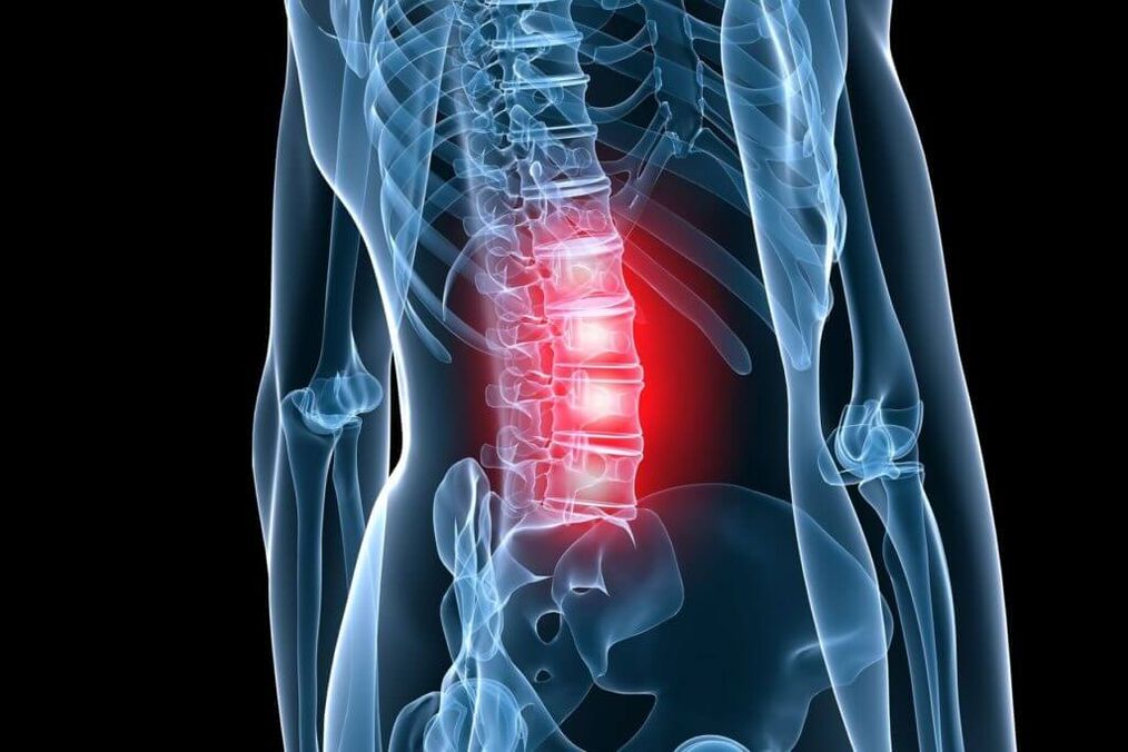
pain4 out of 5 people experience back pain at least once in their lives. They are for the working age populationthe most common cause of disabilitywhich determines their social and economic importance in all countries of the world. Among the diseases that are accompanied by pain in the lumbar spine and limbs, one of the main places is occupied by osteochondrosis.
Osteochondrosis of the spine (OP) is its degenerative-dystrophic lesion, starting from the nucleus pulposus of the intervertebral disc, extending to the fibrous ring and other elements of the spinal segment with frequent secondary effects on neighboring neurovascular formations. Under the influence of unfavorable static-dynamic loads, the elastic pulpous (gelatinous) core loses its physiological properties - it dries up and becomes sequestered over time. Under the influence of mechanical loads, the fibrous ring of the disc, which has lost its elasticity, protrudes, and after that fragments of the nucleus pulposus fall out through its cracks. This leads to the appearance of acute pain (lumbago), because. peripheral parts of the annulus fibrosus contain Luschka nerve receptors.
Stages of osteochondrosis
The intradiscal pathological process corresponds to stage 1 (period) (OP) according to the classification proposed by Ya. Yu. Popelyansky and A. I. Osna. In the second period, not only the cushioning ability is lost, but also the fixing function with the development of hypermobility (or instability). In the third period, the formation of disc herniation (protrusion) is observed. According to the degree of their prolapse, disc herniations are divided intoelastic protrusionwhen there is a uniform protrusion of the intervertebral disc, andsequestered bulge, characterized by uneven and incomplete rupture of the fibrous ring. The nucleus pulposus moves into these rupture sites, creating local protrusions. With a partially prolapsed disc herniation, all layers of the fibrous ring rupture, and possibly the posterior longitudinal ligament, but the hernial protrusion itself has not yet lost contact with the central part of the nucleus. A fully prolapsed disc herniation means that not its individual fragments prolapse, but the entire nucleus into the lumen of the spinal canal. According to the diameter, disc herniations are divided into foraminal, posterolateral, paramedial and medial. Clinical manifestations of disc herniation are different, but various compression syndromes often develop at this stage.
Over time, the pathological process can move to other parts of the spinal movement segment. An increase in the load on the vertebral bodies leads to the development of subchondral sclerosis (hardening), then the body increases the support area due to marginal bony growths around the entire perimeter. Overloading the joint leads to spondylarthrosis, which can cause compression of neurovascular formations in the intervertebral foramen. It is precisely these changes that are observed in the fourth period (stage) (OP), when there is a total lesion of the spinal movement segment.
Any schematization of such a complex, clinically diverse disease as OP is, of course, rather arbitrary. However, it enables the analysis of clinical manifestations in their dependence on morphological changes, which enables not only the establishment of a correct diagnosis, but also the determination of specific therapeutic measures.
Depending on which nerve formations are pathologically affected by disc herniation, bony growths and other affected structures of the spine, reflex and compression syndromes are distinguished.
Lumbar osteochondrosis syndromes
Thatcompressioninclude syndromes in which the root, vessel, or spinal cord is stretched, compressed, and deformed over designated vertebral structures. Thatreflexinclude syndromes caused by the action of these structures on the receptors that innervate them, mainly on the endings of recurrent spinal nerves (Lushka's sinuvertebral nerve). Impulses propagating along this nerve from the affected spine travel through the posterior root to the posterior horn of the spinal cord. Moving to the front horns, they cause reflex tension (defense) of the innervated muscles -reflex-tonic disorders.. Transferring to the sympathetic centers of the lateral horn at their own or adjacent level, they cause reflex vasomotor or dystrophic disorders. Such neurodystrophic disorders occur primarily in low vascularized tissues (tendons, ligaments) at the points of attachment to bony prominences. Here the tissues undergo defibration, swelling, become painful, especially when stretched and palpated. In some cases, these neurodystrophic disorders cause pain that occurs not only locally, but also at a distance. In the latter case, the pain is reflected, it seems to "shoot" when the diseased area is touched. Such zones are called trigger zones. Myofascial pain syndromes can occur as part of referred spondylogenic pain.. With prolonged tension of the striated muscle, microcirculation is disturbed in some of its areas. Due to hypoxia and edema in muscles, sealing zones are formed in the form of knots and threads (as well as in ligaments). The pain in this case is rarely local, it does not coincide with the innervation zone of certain roots. Reflex-myotonic syndromes include piriformis syndrome and popliteal syndrome, the characteristics of which are covered in detail in numerous manuals.
Thatlocal (local) pain reflex syndromesin lumbar osteochondrosis, lumbago is attributed to the acute development of the disease and lumbago in the subacute or chronic course. An important circumstance is the established fact thatlumbago is a consequence of intradiscal displacement of the nucleus pulposus. As a rule, this is a sharp pain, which often bursts. The patient, as it were, freezes in an uncomfortable position, unable to bend. Attempting to change the position of the body causes an increase in pain. Immobility of the entire lumbar region occurs, flattening of the lordosis, and sometimes scoliosis develops.
In case of lumbago - pain, as a rule, painful, aggravated by movement, with axial load. The lumbar region can be deformed, as in lumbago, but to a lesser extent.
Compression syndromes in lumbar osteochondrosis are also diverse. Among them, radicular compression syndrome, caudal syndrome, lumbosacral discogenic myelopathy syndrome are distinguished.
radicular compression syndromeoften develops due to disc herniation at the L levelIV-LVand LV-Sone, because a herniated disc is more likely to develop at this level. Depending on the type of hernia (foraminal, posterior-lateral, etc. ), one or the other root is affected. As a rule, one level corresponds to a monoradicular lesion. Clinical manifestations of L. root compressionVthey are reduced to the appearance of irritation and prolapse in the corresponding dermatome and to the appearance of hypofunction in the corresponding myotome.
Paresthesia(sensation of numbness, tingling) and shooting pains spread along the outer surface of the thigh, the front surface of the lower leg to the zone of the first finger. Hypalgesia can then appear in the corresponding zone. In the muscles innervated by the roots of LV, especially in the front parts of the lower leg, hypotrophy and weakness develop. First of all, weakness is detected in the long extensor of the affected finger - in the muscle that innervates only the root of L.V. Tendon reflexes with an isolated lesion of this root remain normal.
When compressing the spine of Soneirritation and loss phenomena develop in the corresponding dermatome, extending to the zone of the fifth finger. Hypotrophy and weakness cover mainly the posterior calf muscles. The Achilles reflex decreases or disappears. Knee jerk is reduced only when the roots of L are affected.2, L3, Lfour. Hypotrophy of the quadriceps, and especially the gluteal muscles, also occurs with pathology of the caudal lumbar discs. Compression-radicular paresthesia and pain worsen when coughing, sneezing. The pain increases with movement in the lower back. There are other clinical symptoms that indicate the development of root compression, their tension. The most common symptom tested isLasegue's symptomwhen there is a sharp increase in pain in the leg when you try to raise it in a straightened state. An unfavorable variant of radicular syndromes of lumbar vertebrogenic compression is compression of the cauda equina, the so-called.caudal syndrome. It most often develops with large prolapsed median disc herniations, when all the roots at this level are compressed. Local diagnosis is carried out on the upper part of the spine. The pains, usually severe, do not spread to one leg, but as a rule to both legs, the loss of sensitivity affects the area of the rider's pants. In addition to severe variants and rapid development of the syndrome, sphincter disorders are added. Caudal lumbar myelopathy occurs as a result of occlusion of the lower accessory radiculomedullary artery (often at the root of LV, ) and is manifested by weakness of the peroneal, tibial and gluteal muscle groups, sometimes with segmental sensory disturbances. Often, ischemia develops simultaneously in the epiconus segments (L5-Sone) and cone (S2-S5) spinal cord. In such cases, pelvic disorders are also associated.
In addition to the identified main clinical and neurological manifestations of lumbar osteochondrosis, there are other symptoms that indicate the defeat of this spine. This is especially clearly manifested in the combination of damage to the intervertebral disc on the background of congenital narrowing of the spinal canal, various anomalies in the development of the spine.
Diagnosis of lumbar osteochondrosis
Diagnosis of lumbar osteochondrosisit is based on the clinical picture of the disease and additional examination methods, which include conventional radiography of the lumbar spine, computed tomography (CT), CT myelography, magnetic resonance imaging (MRI). With the introduction of MRI of the spine into clinical practice, the diagnosis of lumbar osteochondrosis (LO) has been significantly improved. Sagittal and horizontal tomographic sections allow you to see the relationship of the affected intervertebral disc with the surrounding tissues, including the assessment of the lumen of the spinal canal. Size, type of herniated disc, which roots are compressed and which structures are determined. It is important to determine the compatibility of the leading clinical syndrome with the level and nature of the lesion. As a rule, patients with compression radicular syndrome have a monoradicular lesion, and the compression of this root is clearly visible on MRI. This is relevant from a surgical point of view because. this defines the operational approach.
Disadvantages of MRI include limitations associated with examining claustrophobic patients, as well as the cost of the study itself. CT is a highly informative diagnostic method, especially in combination with myelography, but it should be remembered that the scan is carried out in the horizontal plane, and therefore the level of the alleged lesion must be clinically determined very accurately. Routine radiography is used as a screening examination and is mandatory in the hospital setting. In functional imaging, instability is best defined. Different anomalies of bone development are clearly visible on spondylograms.
Treatment of lumbar osteochondrosis
In PO, both conservative and surgical treatment is carried out. Atconservative treatmentin osteochondrosis, the following pathological conditions require treatment: orthopedic disorders, pain syndrome, impaired ability to fix the disc, muscle-tonic disorders, circulatory disorders in the roots and spinal cord, nerve conduction disorders, cicatricial adhesive changes, psychosomatic disorders. Methods of conservative treatment (CL) include various orthopedic measures (immobilization, traction of the spine, manual therapy), physiotherapy (therapeutic massage and physiotherapy, acupuncture, electrotherapy), prescription of drugs. Treatment should be complex, staged. Each of the CL methods has its own indications and contraindications, but as a rule it is generalprescribing analgesics, non-steroidal anti-inflammatory drugs(NSAIDs),muscle relaxantsandphysiotherapy.
Analgesic effect is achieved by using diclofenac, paracetamol, tramadol. It has a pronounced analgesic effectdrugscontaining 100 mg diclofenac sodium.
Gradual (long-term) absorption of diclofenac improves the effectiveness of the therapy, prevents possible gastrotoxic effects and makes the therapy as convenient as possible for the patient (only 1-2 tablets per day).
If necessary, increase the daily dose of diclofenac to 150 mg, additionally prescribe painkillers in the form of short-acting tablets. In milder forms of the disease, when relatively small doses of the drug are sufficient. If painful symptoms prevail at night or in the morning, it is recommended to take the medicine in the evening.
The substance paracetamol is inferior in terms of its analgesic effect compared to other NSAIDs, so a drug was developed that, along with paracetamol, includes another non-opioid analgesic, propyphenazone, as well as codeine and caffeine. In patients with ischalgia, when caffeine is used, muscle relaxation, reduction of anxiety and depression is noted. Good results were recorded when using the drug in the clinic to relieve acute pain in myofascial, myotonic and radicular syndromes. According to researchers, with short-term use, the drug is well tolerated, practically does not cause side effects.
NSAIDs are the most commonly used medications for PO. NSAIDs have anti-inflammatory, analgesic and antipyretic effects associated with the suppression of cyclooxygenase (COX-1 and COX-2) - the enzyme that regulates the conversion of arachidonic acid into prostaglandins, prostacyclin, thromboxane. Treatment should always start with the appointment of the safest drugs (diclofenac, ketoprofen) in the smallest effective dose (side effects depend on the dose). In elderly patients and patients with risk factors for side effects, it is advisable to start treatment with meloxicam, especially celecoxib or diclofenac/misoprostol. Alternative methods of administration (parenteral, rectal) do not prevent gastroenterological and other side effects. The combined drug diclofenac and misoprostol has certain advantages over standard NSAIDs, which reduces the risk of COX-dependent side effects. In addition, misoprostol can enhance the analgesic effect of diclofenac.
To eliminate pain associated with increased muscle tone, it is recommended to include central muscle relaxants in the complex therapy:tizanidine2-4 mg 3-4 times a day or tolperisone inside 50-100 mg 3 times a day, or tolperisone intramuscularly 100 mg 2 times a day. The mechanism of action of the drug with these substances is significantly different from the mechanisms of action of other drugs used to reduce increased muscle tone. Therefore, it is used in situations where there is no antispastic effect of other drugs (in so-called unresponsive cases). The advantage over other muscle relaxant drugs used for the same indications is that there is no reduction in muscle strength against the background of the decrease in muscle tone. The drug is an imidazole derivative, its action is related to the stimulation of central a2-adrenergic receptors. It selectively inhibits the polysynaptic component of the stretch reflex, has an independent antinociceptive and mild anti-inflammatory effect. The substance tizanidine acts on spasticity of the spine and brain, reduces stretch reflexes and painful muscle spasms. It reduces resistance to passive movements, reduces spasms and clonic convulsions and increases the strength of voluntary contractions of skeletal muscles. It also has a gastroprotective property, which determines its use in combination with NSAIDs. The drug has practically no side effects.
Operationwith PO, it is carried out with the development of compression syndromes. It should be noted that the presence of the fact of disc herniation during MRI is not sufficient for the final decision on surgery. Up to 85% of patients with disc herniation among patients with radicular symptoms pass without surgery after conservative treatment. CL, with the exception of a number of situations, should be the first step in helping patients with PO. If complex CL is ineffective (within 2-3 weeks), surgical treatment (CL) is indicated in patients with disc herniation and radicular symptoms.
There are emergency indications for PO. This includes the development of caudal syndrome, as a rule, with complete prolapse of the disc into the lumen of the spinal canal, the development of acute radiculomyeloischemia and pronounced hyperalgic syndrome, when even the appointment of opioids, blockade does not reduce pain. It should be noted that the absolute size of the disc herniation is not decisive for the final decision on surgery and should be taken into account in connection with the clinical picture, the specific situation observed in the spinal canal according to tomography (for example, there may be a combination of a small hernia on the background of stenosis of the spinal canal orvice versa - the hernia is large, but of medium position on the background of a wide spinal canal).
Open access to the spinal canal is used in 95% of disc herniation cases. Various discopuncture techniques have not been widely used to date, although a number of authors state their effectiveness. The operation is performed with conventional and microsurgical instruments (with optical magnification). During the approach, the removal of bone formations of the vertebrae is avoided by using a mostly interlaminar approach. However, in the case of a narrow canal, hypertrophy of the joint processes, fixed medial disc herniation, it is advisable to expand the access to the bone structures.
The results of surgical treatment largely depend on the experience of the surgeon and the correctness of the indications for a particular operation. According to the convenient expression of the famous neurosurgeon J. Brotchi, who performed more than a thousand osteochondrosis operations, it is necessary to "not forget that the surgeon must operate on the patient, not the tomographic image".
In conclusion, I would like to emphasize once again the need for a detailed clinical examination and tomogram analysis in order to make an optimal decision on the choice of treatment tactics for a particular patient.


